The Role of Technology in Orthodontics
At our orthodontic clinic in Sturbridge, dental technology is used in nearly every aspect of planning and treatment.
We use advanced technology to gain a detailed understanding of your unique orthodontic needs. Our digital scans allow us to provide an accurate diagnosis, leading to highly personalized treatment plans that align with your goals in the most efficient and effective way possible.
Our technology guides us in developing these treatment plans and adjusting them along the way as needed – because your comfort and results matter to us.
From your complimentary consultation to your final appointment, you'll experience how our approach makes orthodontic care simple, comfortable, and precise.
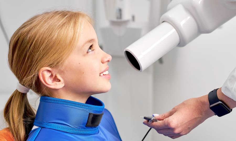
Digital X-rays
Digital X-ray technology uses digital sensors and a computer to produce detailed, high-quality internal images of your teeth and gums.
These digital images allow your orthodontist to see the position of your teeth below the gums, as well as the surrounding bones, helping to diagnose your orthodontic condition. They also assist us in identifying factors that could influence your treatment, such as developing tooth decay and previous dental restorations. All of this information is crucial for diagnosis and treatment planning.
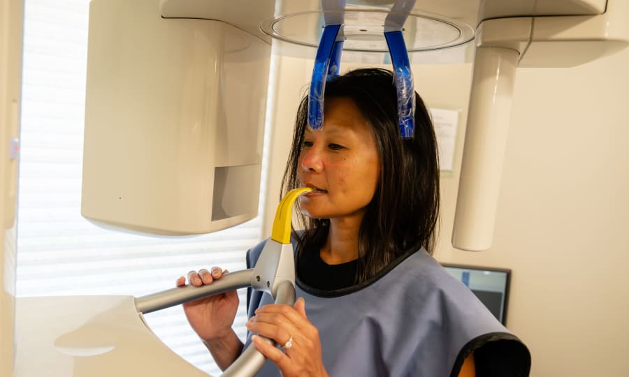
Panoramic X-rays
Panoramic X-ray technology lets your orthodontist take a single, two-dimensional image that shows all of your mouth's oral structures. This includes the teeth, the upper and lower jaws, and the surrounding tissues.
This is a vital tool for assessing the presence or absence of specific teeth and their roots, their form and structure, their eruption sequence, and their relationship to one another in the jaws. This offers valuable insights to help with treatment planning and assessment.
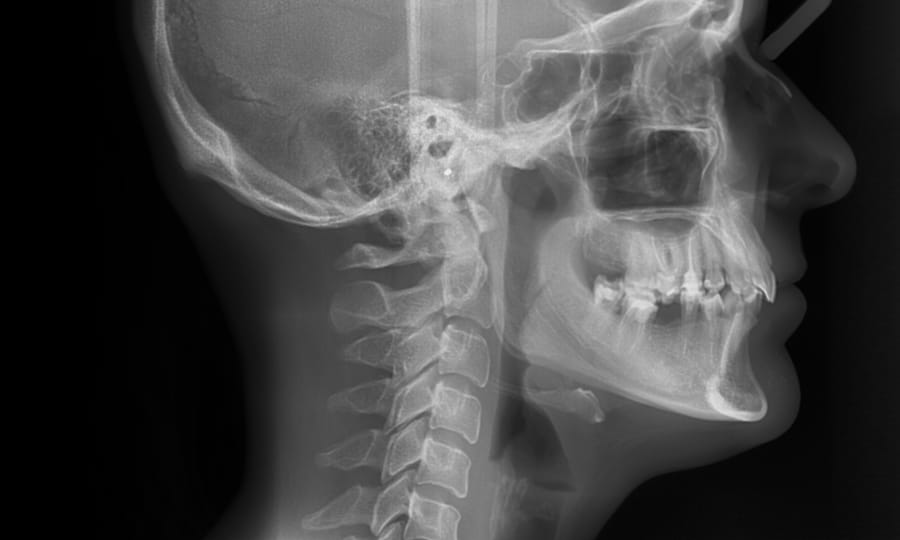
Cephalometric X-rays
A cephalometric X-ray is a specialized image that shows a side view of your head, including your teeth, jaw, and facial profile. This type of imaging helps your orthodontist understand how your teeth and jaw relate to each other and to your facial structure.
Cephalometric X-rays are frequently used to assess jaw growth, identify misalignments, and plan orthodontic treatments such as braces or aligners. They also help monitor changes over time, ensuring your treatment advances as planned and your bite stays in harmony with your facial proportions.
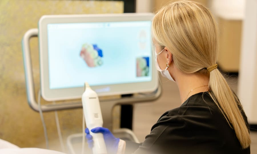
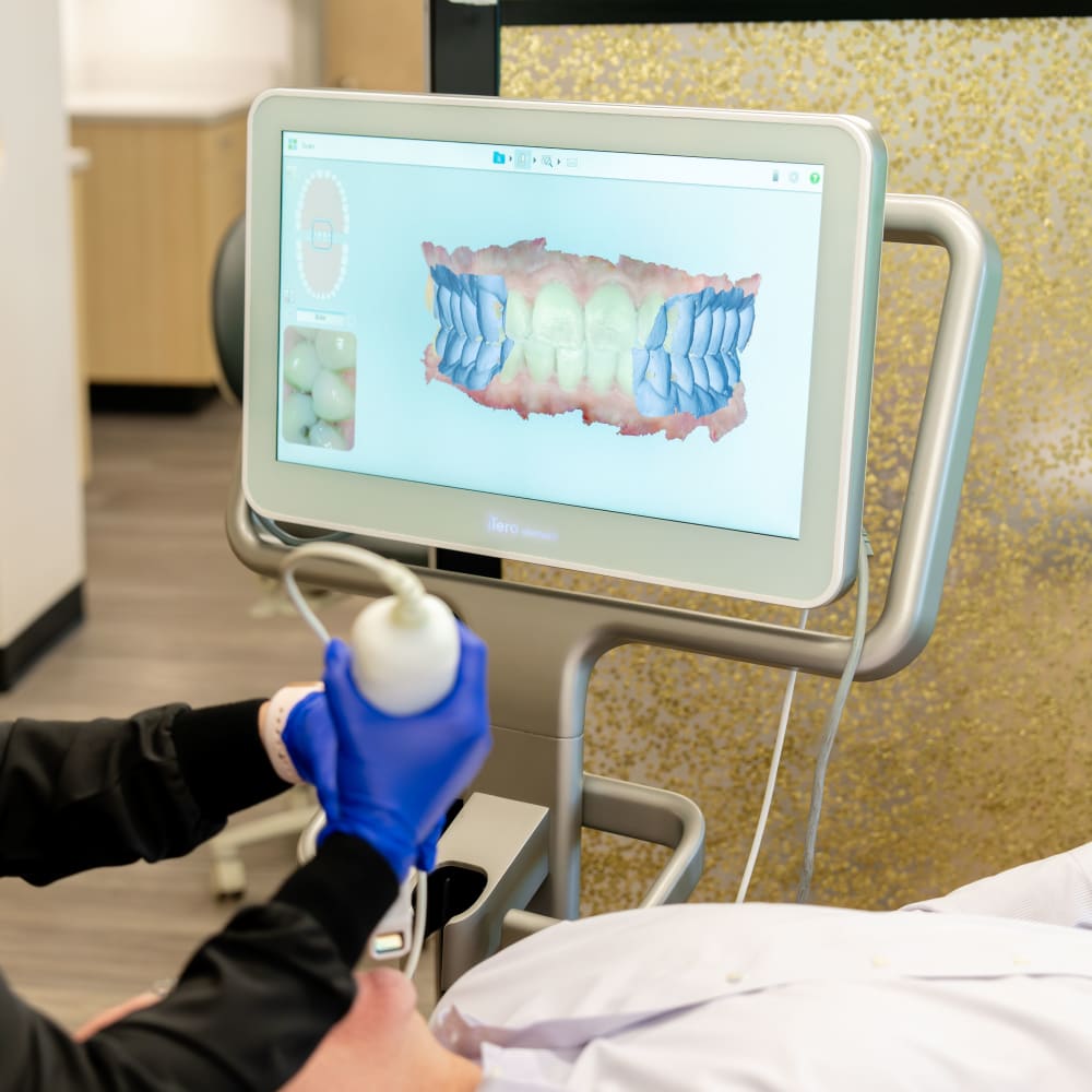
iTero™ Intraoral Scanner
The iTero intraoral scanner is a handheld imaging device that creates highly detailed, three-dimensional digital impressions of your mouth in just minutes.
We simply look inside your mouth as we move the handheld scanner around, and watch as real-time 3D models of your teeth appear on the screen. No mess, no discomfort, just easy, precise imaging.